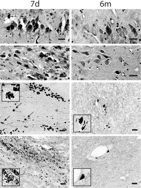
Figure 2.
Representative photomicrographs revealing a modest number of pyknotic neurons in CA1 and CA3 subfields of the hippocampus (top panels) and foamy macrophage infiltrations within subcortical white matter, basal ganglia and lining blood vessels. Axonal bulbs were close to macrophage infiltrations (black arrows), detected by antibody OX42 at 7 days and 6 months post-injury (bottom panels). Bar, 25 μm.

