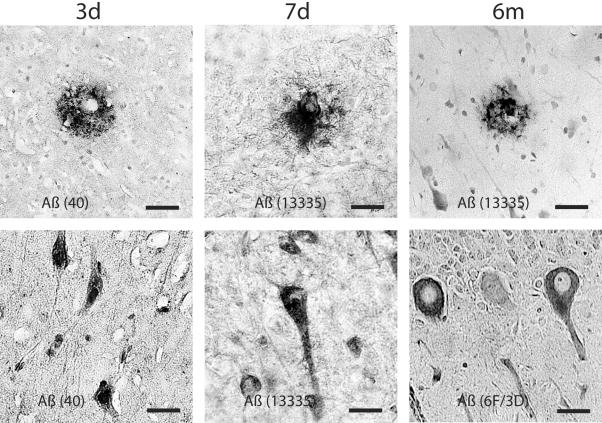
Figure 5.
Representative photomicrographs showing Aβ-containing plaque-like profiles in the gray and white matter. Occasionally, perivascualr Aβ depostis were found (top, middle). Aβ accumulation was also found in cortical and cerebellar neurons at 3 days to 6 months following brain trauma. Aβ was identified by antibodies, 6F/3D, Aβ1–40, 13335. Bar, 25 μm.

