Figure 3.
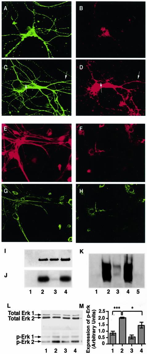
The Mek1,2 inhibitor, U-0126, calpeptin, and Erk1,2 antisense oligonucleotides attenuate Erk1,2 activation and NF-phosphorylation in hippocampal neurons. A–H: Hippocampal neurons (14 DIV) were either untreated (A and B) or treated with 0.5 μmol/L ionomycin (C and D) were immunostained with a monoclonal antibody to β tubulin (A and C) and a polyclonal antibody to phospho-Erk 1,2 (B and D). These neurons were also treated with sense (E and G) and antisense (F and H) oligonucleotides against Erk1,2 mRNA as described in Materials and Methods and were immunostained with a goat polyclonal antibody (Santa Cruz) against Erk1,2 (E and F) and with a monoclonal antibody to phospho-NF-M (G and H). I: Recombinant Erk2 (1 μg) was incubated with the immunprecipitate of Mek1,2, from lysates of ionomycin-treated hippocampal neurons, in the absence (lane 2) and presence (lane 3) of U-0126 (10 μmol/L) or calpeptin (lane 4; 20 μΜ) in a kinase reaction mixture detailed in the text, followed by SDS-PAGE and silver stain. Lane 1 represents the immunoprecipitate of Mek1,2. J: Autoradiogram of Erk2 shown in I. K: Representive image taken of a Western blot for a KSPXXXK fusion protein using the monoclonal antibody RT-97, specific for phosphorylated NF-H and NF-M. The KSPXXXK fusion protein was incubated in a kinase reaction mixture containing the immunoprecipitate of Mek1,2 and recombinant Erk2, in the absence (lane 2) and presence (lane 3) of U-0126 (10 μmol/L) or calpeptin (lane 4; 20 μΜ). Lane 1 represents the mixture of immunoprecipitate of Mek1,2 and Erk2, and lane 5 represents KSPXXXK fusion protein alone. L: Representive image taken of a Western blot for hippocampal neuron cell lysates (15 μg) in vehicle-treated (lane 1), ionomycin (0.5 μmol/L; lane 2), ionomycin and U-0126- (0.5 μmol/L and 10 μmol/L; lane 3) and calpeptin-treated (20 μmol/L; lane 4) neurons for 24 hours, that were probed for Erk1,2 and phospho-Erk1,2 expression. M: Bar graph shows the relative band intensities of p-Erk1,2. Values are expressed as mean ± SEM for three separate experiments. The ionomycin-treated neurons (lane 2) showed a significantly higher activation compared to vehicle-treated controls (lane 1; P < 0.001, n = 3) and the neurons treated with ionomycin together with calpeptin (lane 4; P < 0.05, n = 3).

