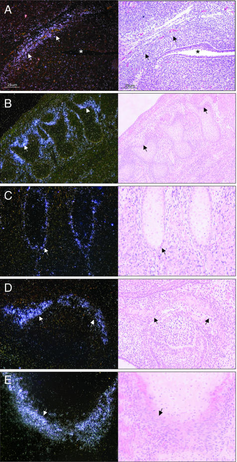
Figure 1.
In situ hybridization of WISP-1 expression during mouse development. Left: Dark-field images; right: corresponding bright-field images. A: Base of the skull dorsal of the oropharynx (*) at E12.5. At E15.5, WISP-1 is expressed in osteoblasts and mesenchymal cells adjacent to bones undergoing endochondral ossification (B, vertebras; C, ribs) and intramembranous ossification (D, ossification within palatal shelf of maxilla). WISP-1 expression was similarly distributed in human embryo lower limb (E, lateral border of head of tibia). Original magnifications: ×100 (A, D); ×40 (B); ×200 (C, E).

