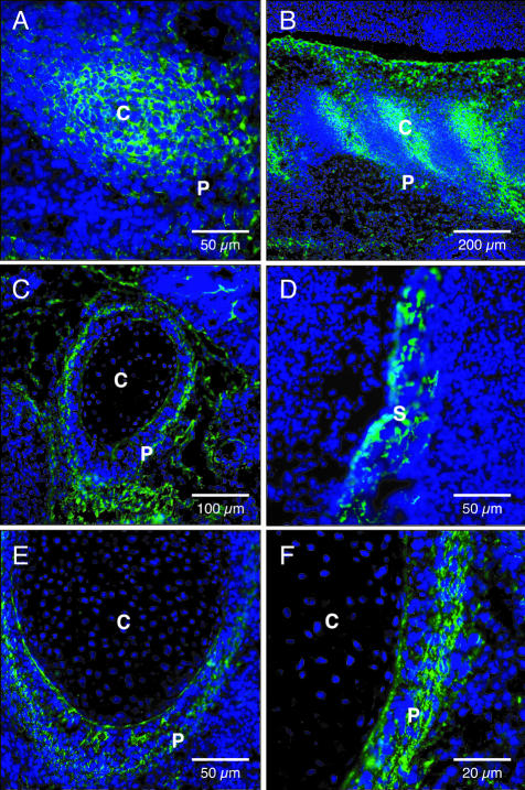
Figure 5.
In situ WISP-1 binding in rat embryo. At E14, WISP-1 binding revealed an intense fluorescent signal associated with costal (A) and vertebral (B) condensed mesenchymal cells. At E18, WISP-1 bound to osteoblasts and perichondral mesenchyme of developing bones; mesenchyme surrounding cartilage primordium of rib (C), calvaria (D), mesenchyme surrounding cartilage primordium of distal part of radius (E, F). P, perichondrium; C, cartilage primordium; S, skull. Original magnifications: ×200 (A, D, E); ×40 (B); ×100 (C); ×400 (F).

