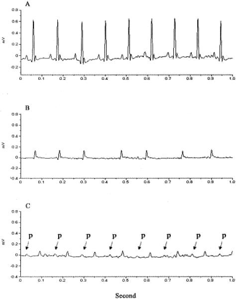
Figure 10.
Ambulatory ECG. ECG was recorded in wild-type and ACE 8/8 mice using telemetry. A: A representative ECG of a wild-type mouse showing normal sinus rhythm. B: A representative ECG from six analyzed ACE 8/8 mice showing low QRS voltage, no P wave, and irregular RR intervals. C: ECG of an ACE 8/8 mouse showing the presence of an atrial rhythm with prolonged AV block. Atrial P waves are labeled with arrows.

