Figure 3.
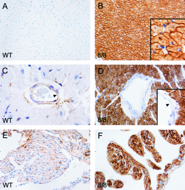
ACE immunohistochemistry of heart. Tissues from wild-type (WT) and ACE 8/8 mice were prepared and stained with an anti-ACE antibody. A and B: Sections of cardiac ventricles. The high-power inset in B shows abundant ACE within the cell membranes of myocytes. Despite this, the overall histological appearance of the ACE 8/8 ventricular myocardium was unchanged from that of wild-type tissue. C and D: High-power sections of the heart showing ventricular blood vessels. Wild-type mice have both endothelial and adventitial expression of ACE. In contrast, blood vessels in the hearts of ACE 8/8 mice have no endothelial expression of ACE. E and F: Sections of atria. As in the ventricles, the atria of ACE 8/8 mice make abundant ACE.

