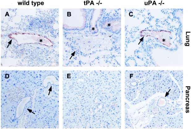
Figure 1-4244.
Immunostaining for tPA in normal lung and pancreas tissues from wild-type (A and D), tPA −/− (B, E) and uPA −/− mice (C and F). Anti-tPA antibodies show a strong reactivity with vascular endothelial cells in the lung of wild-type (A) and uPA−/− (C) mice but not in cells from tPA−/− (B) mice. In the pancreas, tPA is undetectable in normal acinar and ductal cells (D and F, arrows). Asterisk indicates non-specific staining. Original magnification, ×200 (A–F).

