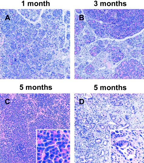
Figure 4-4244.
Histological analysis of Ela1-myc tPA−/− transgenic mice-derived tumors. Sections of paraffin-embeded pancreatic tissue from Ela1-myc tPA−/− transgenic mice were stained with hematoxylin and eosin. The pancreas from young mice shows widespread acinar dysplasia (A and B) whereas tumors in 5- month-old mice display areas of acinar (C) as well as ductal (D) differentiation. C and D and the corresponding insets display different areas from the same tumor. Original magnification; ×100 (A–D), inset, ×400 (C) and ×200 (D).

