Figure 2-4260.
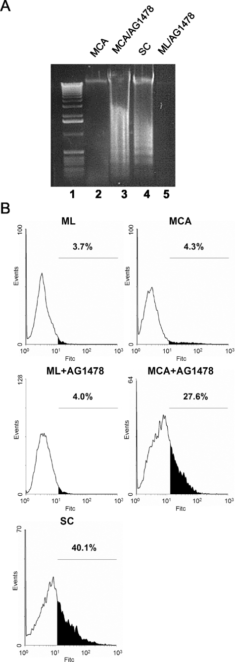
Cell-cell adhesion-induced EGFR activation suppresses anoikis. A: DNA fragmentation of HSC-3 cells. HSC-3 cells were plated as MCA culture in the absence or presence of 1 μg/ml AG1478 for 48 hours (lanes 2 and 3). Suspended single cells (SC) alone (lane 4) and monolayers (ML) cultured for 48 hours in the presence of 1 μmol/L AG1478 (lane 5) were used as controls. The DNA laddering assay for intranucleosomal DNA cleavage was performed as described in Experimental Procedures. Lane 1: Standard 100-bp DNA ladder. B: TUNEL analysis of AG1478-treated HSC-3 cells. HSC-3 cells were plated as ML, MCA, or SC culture for 48 hours before TUNEL analysis. AG1478 (1 μmol/L) was added to the culture as indicated. Values represent the apoptotic cell fractions (%) that stained positive with FITC-dUTP.

