Figure 4.
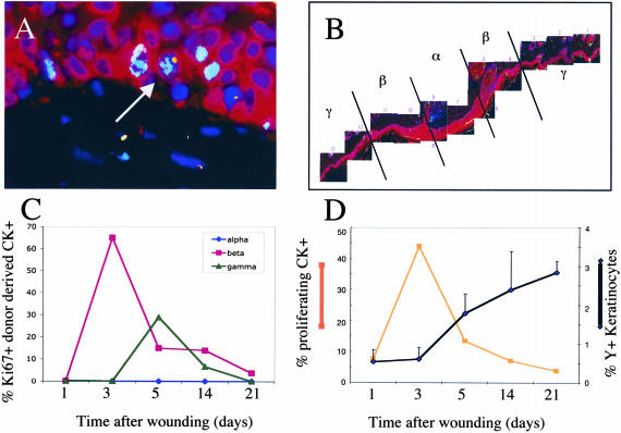
A: Fluorescent staining for Ki67 (light blue nuclei), Y chromosome (yellow), cytokeratin (red), and DAPI for the nuclei (dark blue). The arrow indicates Y chromosome+, Ki67+, and cytokeratin+ cells in the basal layer of the skin in the β region 5 days after wounding. B: Shown is a composite of a skin wound 21 days after wounding. The α region is the center of the wounded area, and the β region is located at the periphery of the wound. Just outside the highly proliferative β region is the γ α region. C: Graph shows the percentage of donor-derived epithelial cells that are Ki67+ in each of the regions delineated in B at different times after wounding (α, circles; β, squares; γ, triangles). D: Graph shows the percentage of all donor-derived epithelial cells that are proliferating over time (squares) on the left axis and the percentage of keratinocytes that are donor-derived (diamonds) on the right axis at different times after wounding.

