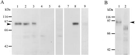
Figure 5.
Immunoblotting of ADAM12m in glioblastoma tissues. A: Homogenates (20 μg/lane) from glioblastoma (lanes 1 to 3 and 6) and nonneoplastic brain tissues (lanes 4 and 5) and cell lysates (20 μg/lane) of CaR-1 (lane 7, a negative control) and U251 (lane 8, a positive control; lane 9, absorption test) were resolved by sodium dodecyl sulfate-polyacrylamide gel electrophoresis. The proteins in the gels were transferred onto polyvinylidene difluoride membranes and the membranes immunostained with anti-ADAM12m antibody (2 μg/ml) (lanes 1 to 5, 7, and 8), the antibody (2 μg/ml) absorbed with antigen peptide (3 μg/ml) (lane 9), or nonimmune mouse IgG (2 μg/ml) (lane 6) as described in Materials and Methods. Note that a positive band of ∼90 kd is found in the glioblastoma samples (lanes 1 to 3) and the U251 cell lysates (lane 8), whereas no such species is recognized in the nonneoplastic brain samples (lanes 4 and 5) or CaR-1 cell lysates (lane 7). Only a background staining is present with nonimmune IgG (lane 6), and a negligible band is observed with the antibody absorbed with the peptide (lane 9). B: Deglycosylation of ADAM12m. Homogenates were treated with N-glycosidase F and subjected to immunoblotting for ADAM12m as described in Materials and Methods.

