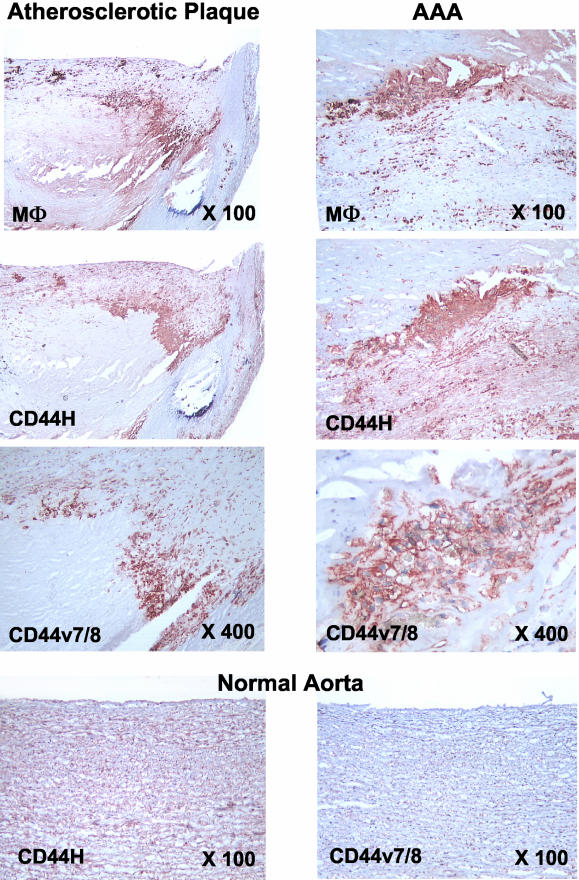
Figure 2.
Increased CD44 expression in macrophage-rich areas in atheroma and AAA. Both CD44 and its splice variants are prominently expressed in macrophage-rich areas of atherosclerotic plaques (left) and AAA tissue (right). Representative stainings are shown. Staining of tissue samples from seven patients showed similar results. Normal, nondiseased, aorta (bottom, n = 5) is positive for CD44H but not CD44v7/8. Original magnifications are indicated.

