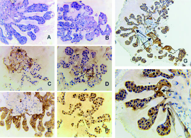
Figure 4.
Formalin-fixed, paraffin-embedded sections of EIII8-HUVEC three-dimensional co-cultures were either stained with H&E (A and B) or with antibodies to cd31 (C), factor VIII (D), cytokeratins (E), proliferating cell nuclear antigen (F), or galectin-3 monoclonal TIB-166 antibody (G and H). Note the widespread immunoreactivity to cytokeratins in the branching end buds as opposed to the localized cd31 and factor VIII8-expressing endothelial cells. Also note the presence of numerous proliferating cells in branching end buds invading into the surrounding ECM. Note that galectin-3 staining is exclusively localized in epithelial buds with higher TIB-166 immunoreactivities in epithelial cells that are adjacent to endothelial cells (arrows, G and H). Original magnifications: ×25 (A–F); ×40 (H); ×10 (G).

