Figure 5.
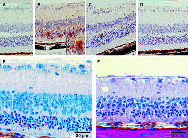
Apoptosis in retinas of scrapie-infected transgenic mice. Shown are representative retinal sections stained by TUNEL of mock-infected tg7 mice (A) and scrapie-infected tg7 (B), tgNSE (C), and tgGFAP (D) mice. tgGFAP and tgNSE retinas were stained at the clinical time of disease, and tg7 retinas were stained midway through disease (see Figure 3 legend for precise times). B: Characteristic pyknotic nuclei from apoptotic cells in tg7 retinas are indicated by white arrowheads (inset). E: Staining with anti-cleaved caspase-3 at a higher magnification shows typical cytoplasmic staining (white arrows). F: In a different mouse with more severe retinal degeneration, H&E staining shows retinal atrophy and cell loss in outer nuclear layer with typical pyknotic nuclei and apoptotic bodies (white arrows). Original magnifications, ×40.

