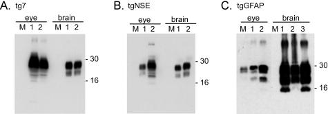
Figure 6.
Western blot of PrP-res from clinically sick transgenic mice. Shown are representative blots from tg7 (A), tgNSE (B), and tgGFAP (C) eye and brain tissue at the clinical time of infection after intraocular inoculation with hamster scrapie. Each 10% (w/v) homogenate was treated with proteinase K as described in the Materials and Methods, and PrP-res bands representing multiple glycosylation forms are indicated by approximate molecular weight markers (16 to 30 kd). M (mock), uninfected brain or eye homogenate. Lanes 1 to 3 represent protein from up to three individual animals. All blots were loaded with an equivalent amount of protein (5 mg/10 μl tissue equivalents). All exposure times were equivalent except for tgGFAP eye, which was 100-fold longer because of low levels of detectable PrP-res. This very long exposure also resulted in visualization of a band in the lane with mock-infected eye. It is unclear whether this is a non-PrP protein or actual PrP-res from the adjacent lane.

