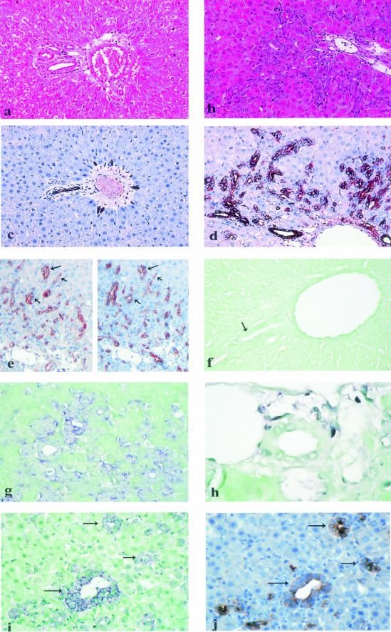
Figure 1.
Histopathological findings and stromal cell-derived factor-1 (SDF-1) protein and mRNA expression in normal liver and in hepatic regeneration from oval cells in rat [2-acetylaminofluorene (AAF)/partial hepatectomy (PH) model]. a: Normal liver (hematein-eosin stain, original magnification, ×200). b: Liver from AAF-treated rats (day 9 after PH); there is marked periportal oval cell accumulation (hematein-eosin stain, original magnification, ×400). c: Normal liver (same portal area as in Figure 1a); SDF-1 protein is immunohistochemically detected in interlobular bile duct epithelial cells and in rare periportal ductules (original magnification, ×200). d: Liver from AAF-treated rats (day 9 after PH); SDF-1 protein is strongly expressed by oval cells forming periportal ductular structures, with increased labeling at the periphery of the cell cytoplasm; SDF-1 protein is also expressed by interlobular bile duct epithelial cells (original magnification, ×400). e: Liver from AAF-treated rats (day 9 after PH); on serial sections, the same ductules (arrows), made of oval cells, express alphafetoprotein (left) as well as SDF-1 protein (right). (f) Portal area in a normal liver (an interlobular bile duct is indicated by an arrow); absence of SDF-1 mRNA detection with an antisense probe (original magnification, ×400). g: Liver from AAF-treated rats (day 9 after PH); SDF-1 mRNA is demonstrated by in situ hybridization with an antisense probe in periportal oval cells (original magnification, ×400). h: Liver from AAF-treated rats (day 9 after PH); absence of SDF-1 mRNA detection in an interlobular bile duct (original magnification, ×1000). i and j: Serial sections of a liver from AAF-treated rats (day 9 after PH); SDF-1 mRNA (i) as well as alphafetoprotein (j) are detected in the same ductules (arrows), made of oval cells.

