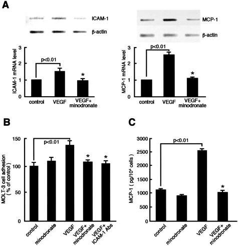
Figure 5.
Effects of minodronate on ICAM-1 and MCP-1 expression in VEGF-exposed ECs. A: ECs were incubated with or without 10 ng/ml of VEGF for 4 hours in the presence or absence of 10 μmol/L minodronate. Thirty ng of poly(A)+ RNAs were transcribed and amplified by PCR. Each bottom panel shows quantitative representation of ICAM-1 and MCP-1 gene induction. Data are normalized by the intensity of β-actin mRNA-derived signals and then related to the value of the control. B: ECs were incubated with or without 10 ng/ml of VEGF for 24 hours in the presence or absence of 10 μmol/L minodronate or 1 μg/ml of mAbs against human ICAM-1. Molt-3 cell adhesion was measured as fluorescent intensity. The percentage of Molt-3 cell adhesion is related to the value of control cells. C: MCP-1 content in the medium was measured. *, P < 0.01 compared to the value with VEGF alone (n = 4 to 6 per group). Similar results were obtained in two independent experiments.

