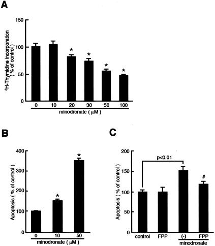
Figure 6.
Effects of minodronate on DNA synthesis (A) and apoptosis (B and C) in cultured melanoma cells. G361 melanoma cells were incubated with the indicated concentrations of minodronate in the presence or absence of 0.5 μg/ml of FPP for 24 hours. The percentage of [3H]thymidine incorporation is related to the value of the control. One hundred percent indicates 38,620 cpm. Apoptotic cell death was measured as absorbance at 405 nm. The percentage of apoptotic cell death is related to the value of the control without minodronate. *, P < 0.01 compared to the control value. #, P < 0.01 compared to the value with 10 μmol/L minodronate alone. N = 4 to 6 per group. Similar results were obtained in two independent experiments.

