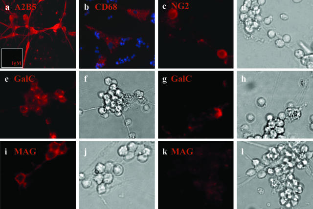
Figure 1.
Immunocytochemical characterization of the A2B5+ cell fraction derived from the adult human CNS. After magnetic bead selection, A2B5+ cells derived from human adult CNS were grown in DMEM F12 supplemented with N1, bFGF, and T3, kept in culture for 7 days, and then immunostained for different neural markers; a: A2B5+ cells (red) with a small inset for the isotype control IgM (red); b: CD68+ cells (red) with nuclei double-stained with Hoechst (blue); c and d: NG2+ cells (red) and corresponding bright field; e–h: GalC+ cells (red) with corresponding bright fields; i–l: MAG+ cells (red) and bright fields. Both for GalC and MAG two different fields are shown to represent cell aggregates that are mostly positive (e and i) or mostly negative (g and k) for GalC and MAG. Original magnifications: ×40 (a, c–l); ×20 (b).

