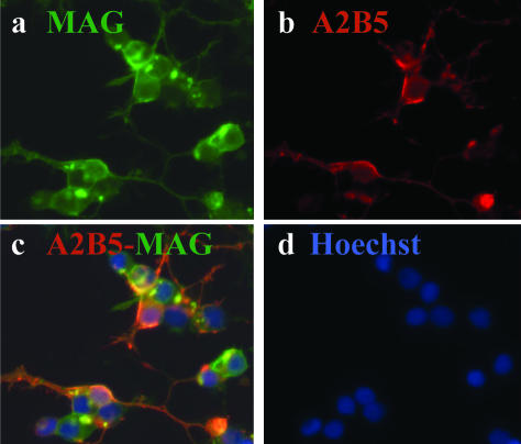
Figure 2.
Immunocytochemical characterization of adult OLGs indicating that a fraction of these cells co-express A2B5. After 1 week in culture in MEM supplemented with 5% fetal calf serum, OLGs were co-stained for the myelin antigen MAG and for A2B5. Each staining is shown individually as well as the overlay: a: MAG+ cells (green); b: A2B5+ cells in red; c: overlay indicating co-staining; d: nuclear stain Hoechst for the same field. Original magnifications, ×40.

