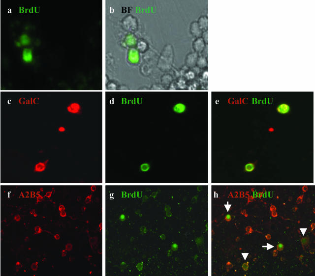
Figure 6.
Comparison of BrdU labeling of adult and fetal A2B5+ cell fractions. A2B5+ cells from adult and fetal material were incubated for 48 hours with BrdU and then cultured for 5 to 6 additional days before staining for BrdU and neural markers. a and b: Adult A2B5+-positive cell fraction stained for BrdU (a) with the respective bright field (b). c–e: Double staining of adult A2B5+ cells for BrdU and GalC. Single stains are shown (c, GalC; d, BrdU) as well as overlay of the two stains (e), indicating co-localization. f–h: Fetal-derived A2B5+ cells co-stained for A2B5 (f) and BrdU (g); overlay of stains is shown in h. Arrowheads show weakly positive cells; long arrows show cells that are strongly positive. Original magnifications: ×60 (a–e); ×40 (f–h).

