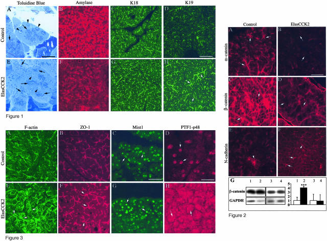
Figure 2.
Immunohistochemistry for adhesion proteins on cryostat sections of wild-type and ElasCCK2 pancreas using antibodies against adherens junction proteins. A and B: α-Catenin, C and D: β-catenin, and E and F: N-cadherin. In ElasCCK2 exocrine pancreas expression of α-catenin and β-catenin at cell-cell contacts is strongly reduced (arrows in B and D) and some residual β-catenin diffusively localized (arrows in D), whereas N-cadherin staining at acinar cell-cell contacts is increased in ElasCCK2 exocrine pancreas (arrows in F). The arrowhead in E points to cell borders between pancreatic islet cells, which normally are strongly positive for N-cadherin in wild-type as well as in ElasCCK2 mice. G: Western blot analysis from soluble (lanes 1 and 2) and total (lanes 3 and 4) proteins of control (lanes 1 and 3) and of ElasCCK2 acini (lanes 2 and 4) and corresponding densitometric analysis of data. Western blot studies were performed using an anti-β-catenin antibody and, to normalize to equivalent protein amount, the blot was reprobed with an anti-GAPDH antibody. One Western blot experiment representative of four is shown. Results of quantification represent means ± SEM of four independent experiments and are expressed as fold control with control expression set to one. White bars, control; black bars, ElasCCK2. ***, Significance at P < 0.001 determined using the Student’s t-test. Scale bar, 60 μm.

