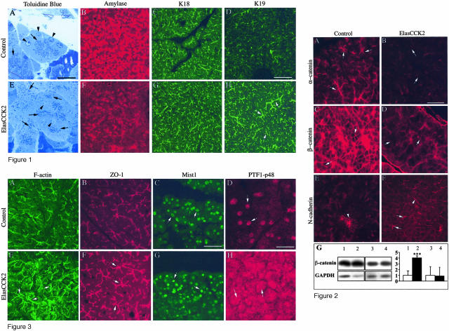
Figure 3.
Immunohistochemistry of cell junctional components and acinar transcription factors on cryostat sections of wild-type and ElasCCK2 pancreas. A–H: Sections are stained with Alexa 488-phalloidin to detect F-actin (A, E), and with antibodies against ZO-1 (B, F), Mist1 (C, G), and PTF1-p48 (D, H). In ElasCCK2 exocrine pancreas, enhanced F-actin staining (compare E with A) as well as enlarged F-actin- and ZO-1-positive duct-like structures (arrows in E and F) are observed. Although Mist1 expression and localization is unaffected in ElasCCK2 mice (arrows in G), a striking amount of PTF1-p48 is seen in the cytoplasm, besides its normal nuclear localization, in ElasCCK2 exocrine pancreas (arrows in H). Note nuclear dysplasia in ElasCCK2 pancreas (G). Scale bars: 60 μm (A–C, E–G); 30 μm (D, H).

