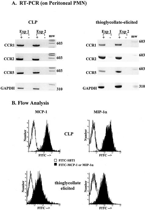
Figure 6.
Expression of CCR 1, 2, 5 and binding of CC chemokines to peritoneal PMN from CLP-operated or thioglycollate-treated mice. A: RT-PCR analysis of CCR1, CCR2, and CCR5 expression in PMN after 6 hours CLP or thioglycollate treatment. Samples were run in the absence (−) and presence (+) of reverse transcriptase to confirm lack of DNA contamination. Blots are from two separate experiments each where n = 4 mice per experiment. B: Binding of PMN to labeled MCP-1 and MIP-1α after 6 hours CLP or thioglycollate treatment. FITC-SBTI was used as a control peptide for binding studies. Histograms are representative of two experiments performed in duplicate with n = 4 mice per experiment.

