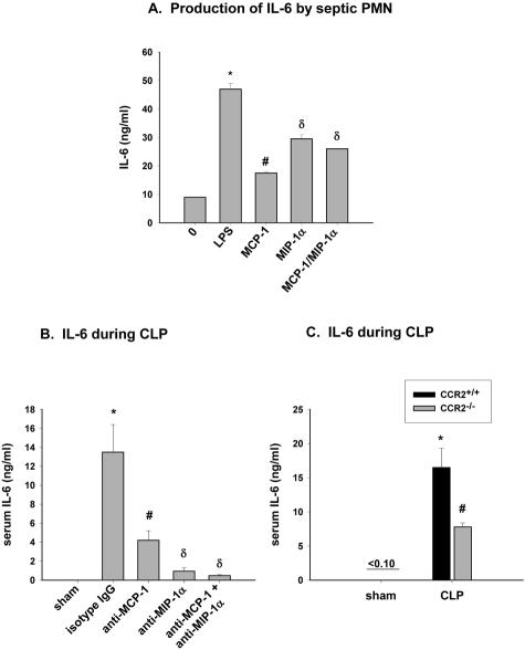
Figure 7.
Role of MCP-1 and MIP-1α on serum IL-6 levels during CLP. A: Peritoneal PMN were isolated from CLP mice 6 hours after CLP and stimulated for 4 hours with either 20 ng/ml LPS (positive control) or 5 ng/ml MCP-1 and MIP-1α, each alone or together, and IL-6 release into supernatant was measured. Values represent mean ± SEM of two experiments performed in triplicate, with n = 4 mice per experiment, where * and # is P < 0.05 compared to no treatment, and δ is P < 0.05 compared to MCP-1-treated. B: Serum IL-6 levels in CLP mice treated with antibodies to MCP-1 and MIP-1α, each alone or together. Six hours after CLP, serum was collected, and IL-6 measured by ELISA. Values represent mean ± SEM of two experiments with n = 4 mice per treatment group, where * and # is P < 0.05 compared to sham-operated and δ is P < 0.05 compared to anti-MCP-1-treated. C: IL-6 was measured in serum from CCR2+/+ and CCR2−/− mice 6 hours after CLP. Values represent the mean ± SEM of two experiments with n = 4 mice per group where * is P < 0.05 compared to sham-operated and # is P < 0.05 compared to CCR2+/+ CLP group.

