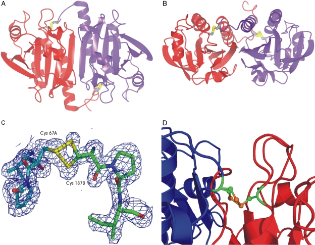
Fig. 3.
A. The PfTrx-Px2 dimer with subunits coloured in purple and red. This figure was made using the program ccp4mg (Potterton et al., 2004).
B. Orthogonal view to A to show the intersubunit disulphide bridges.
C. Electron density in the vicinity of the intermolecular cystine. The 2Fobs − Fcalc, αcalc map is contoured at the 1 σ level. The chains are coloured in cyan and green and the dual conformation of the resolving cysteine from chain B is evident.
D. Ribbon diagram showing the active site disulphide in the context of the Cp loop (red) and the C-terminal tail (blue).

