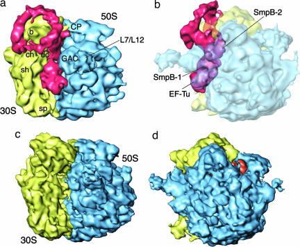
Fig. 2.
Reconstruction of tmRNA–SmpB complex bound with the 70S ribosome and EF-Tu. (a and b) Cryo-EM map generated for 70S·mRNA·tmRNA·EF-Tu·SmpB·S1 in the presence of GTP and kirromycin, in different orientations related by a rotation around the vertical axis. In a, the tmRNA density expands along finger-like protrusions to make contact with the 30S subunit when the density threshold is lowered (data not shown). In b, density for the ribosome is shown semitransparent; density for EF-Tu·tmRNA·SmpB is in red. (c and d) Cryo-EM map of 70S·mRNA·tRNA control. The 50S subunit is in blue, the 30S subunit is in yellow, and the E-site tRNA is in orange. Landmarks on the 50S subunit are as follows: CP, central protuberance; L7/L12, stalk formed by proteins L7/L12. Landmarks on 30S subunit are: sh, shoulder; b, beak; dc, decoding center; ch, entrance of mRNA channel; sp, spur.

