
Fig. 4.
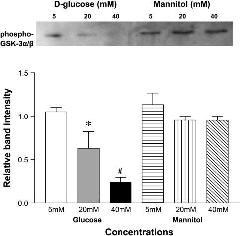
High d-glucose concentration decreases Akt activity in ECs. Akt was immunoprecipitated from HUVEC lysates using immobilized Akt antibody slurry. Immunoprecipitate was incubated with GSK-3 fusion protein in presence of ATP and kinase buffer. Phosphorylation of GSK-3 was used as a measure of Akt activity. Bar graphs show the means relative band intensity ± SE of phospho-GSK-3α/β (Ser 21/9) in lysates. *P < 0.05 and #P < 0.001 compared with control (5 mM d-glucose).