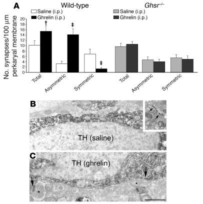
Figure 3. Synaptic remodeling induced by peripheral ghrelin in VTA DA cells.
(A) Ghrelin increased the number of synapses on VTA DA cells of wild-type mice (n = 5). Both total and asymmetric synaptic contacts were elevated, while the number of symmetric synapses was decreased. No synaptic changes were observed after peripheral ghrelin injection in Ghsr–/– mice (n = 5). †P < 0.05, ‡P < 0.01 versus respective saline-treated controls. (B and C) Electron micrographs showing typical TH-immunoreactive perikarya of the VTA from saline- (B) and ghrelin-treated (C) wild-type mice. Arrows in inset of B indicate a symmetric synapse. Arrows in C indicate asymmetric synaptic contacts. Asterisks indicate unlabeled axon terminals. Scale bar: 1 μm.

