
FIGURE 5. Intracellular colocalization of opsin with calreticulin, an ER marker.
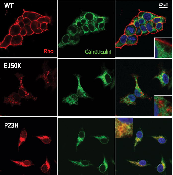
HEK293 cells expressing WT (top row), E150K (middle row), or P23H (bottom row) opsin are stained with both anti-Rho 1D4 (red) and anti-calreticulin (green). The nuclei are stained by Hoechst 33342 (blue). The right column shows the merged images. The insets in the right column are high magnification images of the merged images. The scale bar represents 20 μ m.