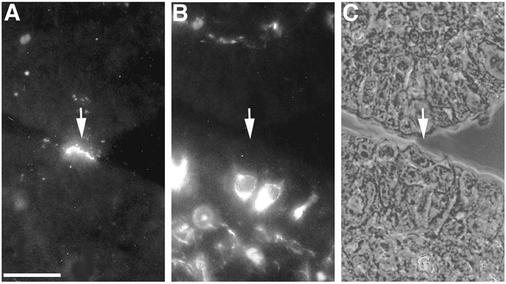
FIG. 1.
Reovirus T1L adhered to M cell apical surfaces in an overlay assay on sections of a rabbit Peyer's patch. Deparaffinized 5-μm sections of Peyer's patch mucosa were dual labeled with biotinylated T1L ISVPs and an anti-vimentin MAb. Bound ISVPs were visualized with streptavidin-TRITC, and vimentin was visualized with an FITC-conjugated secondary antibody. Sections were viewed by fluorescence (A and B) and phase-contrast (C) microscopy. ISVPs (A) (arrow) adhered to apical surfaces of most (but not all) of the epithelial cells that were identified as M cells by dual labeling with the MAb specific for vimentin, an intermediate filament protein that is concentrated around the nuclei of M cells (B). A phase-contrast image of the same section (C) confirmed that the virus-positive cell shows M cell features. Bar, 25 μm.

