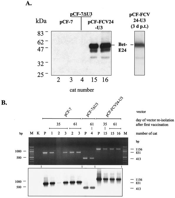
FIG. 6.
Immunoblotting and PCR analysis of FFV-based vectors reisolated from PBLs of vaccinated cats. (A) Detection of Bet-E24 fusion proteins expressed from reisolated pCF-FCV24 vectors. CRFK cell extracts transduced with vectors pCF-7 (from cats 2 and 3), pCF-7ΔU3 (from cat 4), and pCF-FCV24 (from cats 15 and 16) reisolated 61 days after the first vaccination were analyzed by immunoblotting with FCV KS20 antiserum. The position of the authentic Bet-E24 protein expressed by reisolated pCF-FCV24 vectors is marked and compared to that in an extract of pCF-FCV24-transfected CRFK cells 3 days post transfectionem (3 d p.t.) (single lane to the right). The positions of marker proteins are given in the left margin. (B) PCR analysis of the bel-LTR region of reisolated vectors. To PCR amplify the part of the vector genomes where the FCV E sequences were inserted and where part of the U3 region was deleted (compare with Fig. 1C), DNA extracts of reisolated FFV-based vectors were used as indicated. The amplicons were analyzed by agarose gel electrophoresis and ethidium bromide staining (top) and subsequent Southern blot hybridization with the probe marked in Fig. 1C. The date of reisolation, the names of the vectors, and the number designations of the cats are indicated above the images. In lanes P, a plasmid control of the corresponding vectors was amplified, and lane K did not contain any template DNA. In lanes M, molecular size markers were separated, and the 1,000- and 500-bp bands are marked in the left margin. In the right margin, the expected sizes of the amplicons from vectors pCF-7 (831 bp), pCF-7ΔU3 (413 bp), and pCF-FCV24 (1,156 bp) are indicated.

