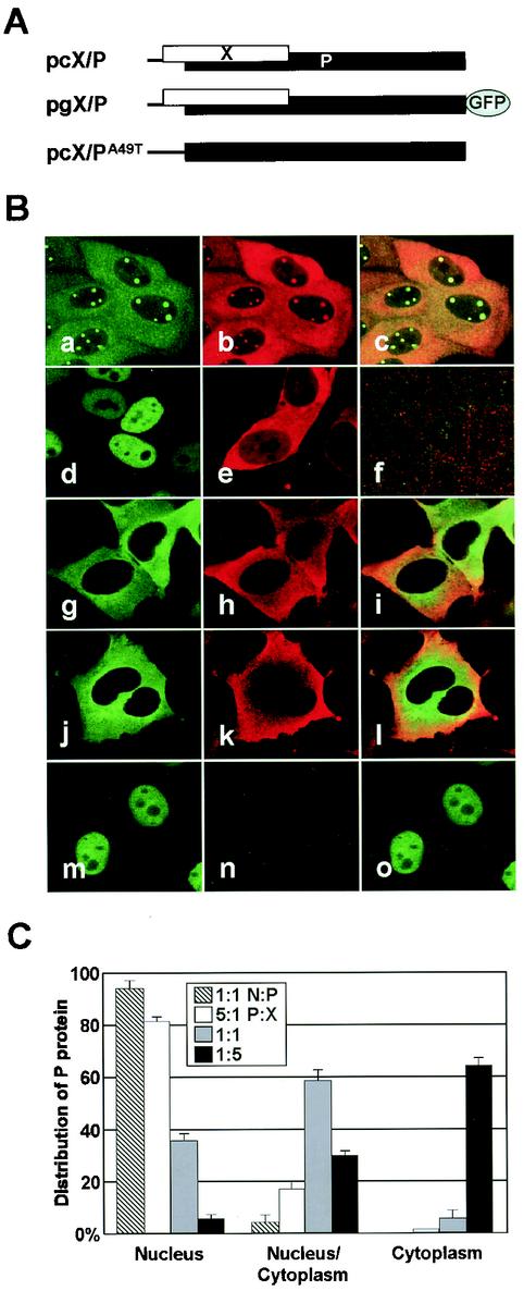
FIG. 1.
Intracellular localization of BDV phosphoprotein. (A) Construction of expression plasmids that contain a BDV cDNA clone corresponding a bicistronic X/P mRNA. (B) Subcellular localization of BDV X and P in infected (panels a to c) and transiently transfected cells. The cells were transfected with expression plasmids as follows: panel d, pcP; e, pcX; f, mock; g to i, pcX/P; j to i, pgX/P; and m to o, pcX/PA49T. The expressions of P and X were detected with anti-P (fluorescein isothiocyanate in panels a, d, g and m) and -X (Cy3 in panels b, e, h, k, and n) antibodies and GFP fluorescence (panel j). The overlap in the distribution of X and P is evident in the merged image (panels c, f, i, l, and o). (C) BDV X promotes cytoplasmic localization of P. The OL cells were cotransfected with pgP and pcX expression plasmids in the ratios of 5:1 (0.5:0.1 μg), 1:1 (0.25:0.25 μg), and 1:5 (0.1:0.5 μg) on eight-well chamber slides. Twenty-four hours posttransfection, the subcellular localization of P was visualized by GFP fluorescence, and the percentage of cells showing each type of P distribution in the transfected cells was determined. P and N expression plasmids (ratio 1:1 [0.25:0.25 μg]) were also cotransfected into OL cells, as control.

