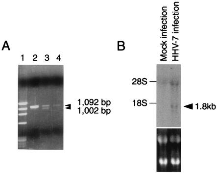
FIG. 1.
(A) Agarose gel (ethidium bromide stained) showing the results of PCR and RT-PCR amplification of HHV-7 DNA and mRNA, obtained from HHV-7-infected cell lines at day 7 postinfection. A 30-cycle PCR amplification was performed on a 1,102-bp fragment from the U12 gene of HHV-7. Lane 1, DNA size markers (φX174/HaeIII digest); lane 2, DNA from infected SupT1 cells; lane 3, cDNA from infected SupT1 cells treated with RT; lane 4, same as lane 3 but without RT. Arrowheads indicate the position of U12 and the splice variant. (B) Northern blot analysis of U12 expression in SupT1 cells infected with HHV-7. Total RNA was isolated from mock-infected and HHV-7-infected SupT1 cells on day 3 after infection. The lower panel shows ethidium bromide staining. The arrowhead indicates the U12 transcript.

