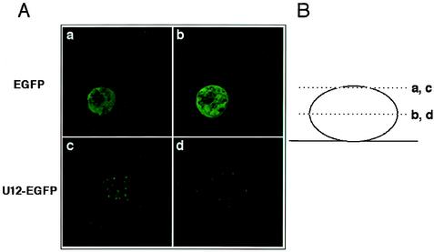
FIG. 4.
Optical sectioning. (A) Confocal laser microscopy analysis and deconvolution software confirmed that K562 cells were transfected with either the pEGFP-N1 vector alone (a and b) or the U12-pEGFP fusion vector (c and d). EGFP products resulted in a predominantly cytoplasmic distribution with a strong signal, but not all cells were labeled. In contrast, the U12-GFP fusion protein accumulated in large granules that were clearly distinct from the more numerous speckles seen on the cell surface. (B) Cells were observed 24 h after transfection in two planes.

