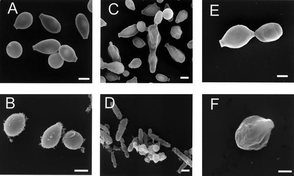
FIG. 1.
Scanning electron micrographs of S. schenckii conidia and yeast cells before and after treatment with enzymes, denaturant, and hot acid. (A and B) Conidia before and after treatment, respectively; (C and D) yeast cells grown in BHI broth before and after treatment, respectively; (E and F) yeast cells grown in minimal medium broth before and after treatment, respectively. Bars, 1 μm.

