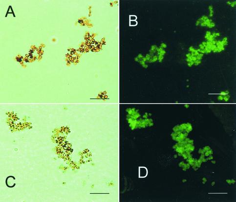
FIG. 4.
Corresponding bright-field (A) and immunofluorescent (B) microscopic images of S. schenckii melanin particles from conidia and corresponding bright-field (C) and immunofluorescence (D) images of yeast cells grown in BHI broth after the preparations were reacted with anti-S. schenckii melanin MAb 7C5 (representative of a novel panel of MAbs). Bars, 10 μm.

