Abstract
The gerHabc operon of Bacillus anthracis, encoding a gerA-like family member of germinant sensors, was shown to be required for endospore germination in the presence of macrophages and in macrophage-conditioned media. The loss of the germination phenotype in macrophage cultures of B. anthracis gerH-null endospores was restored by complementation in trans with a wild-type copy of gerH expressed under the control of its own promoter. Using endospores from both the parental strain B. anthracis Sterne and an isogenic gerH-null strain, we partially characterized germinants secreted by macrophages into the extracellular medium.
Bacillus anthracis endospores can persist for long periods in soil but, upon entering a host, germinate quickly into vegetative bacilli that grow and cause disease (4). Endospores germinate inside of or proximal to phagocytes. These cells are believed to be de facto sites of germination and contribute to the establishment of B. anthracis infection, as phagocytes are the primary means of bacterial access to the body in cases of inhalation anthrax (6, 8, 10, 20). Several recent studies examined the fate of B. anthracis endospores in macrophages (Mφ). Guidi-Rontani et al. showed that murine alveolar phagocytes ingest endospores that then germinate and grow into vegetative bacilli but do not replicate (6). Welkos et al. observed intact endospores and recently germinated nascent bacilli within phagosomes in Mφ that are subsequently destroyed by these immune cells (26, 27, 28). Work by Dixon et al. indicated that newly vegetative bacilli escape from the phagocytic vesicles of the Mφ where they can replicate freely in the host cell cytoplasm and release themselves from the Mφ (3). Though there are important differences among these researchers' observations of the ultimate fates of phagocytosed endospores, it is generally agreed that germinant sensors recognize and bind small-molecule germinants and initiate the germination cascade (17). These sensors belong to the GerA family of germinant receptors, which are best characterized in Bacillus subtilis but which have also been studied in other Bacillus spp. (2, 5, 15, 16, 18, 19, 29, 30).
Six tricistronic gerA-like chromosomal loci (gerAa, gerAb, gerAc) plus the gerX locus carried on pXO1 exist in B. anthracis; these loci encode germinant recognition proteins (6, 10, 11, 25). The gerS operon in B. anthracis mediates germination in response to aromatic ring structures and is required for germination in the presence of cultured Mφ (11, 12). The gerH operon also mediates germination in response to aromatic ring structures and germination triggered by inosine (Ino) in combination with amino acids (25). The germinants recognized by the gerX operon on pXO1 have not yet been identified, although ΔSterne strains lacking pXO1 do not exhibit any gross germination defects in vitro (11, 12). Endospores from a gerX-null strain, identified by Guidi-Rontani et al., germinates slightly less well than do Sterne endospores in the presence of Mφ (6, 12). One ger locus has homology to the gerL locus of Bacillus cereus (alanine response) (1), and another has homology to the gerA locus of B. subtilis (alanine response) (15, 29) but has frameshift mutations in the b and c moieties. The remaining two loci have yet to be investigated.
In the host, germination of B. anthracis endospores constitutes the first stage of establishment during infection. It is essential for B. anthracis to recognize an appropriate host environment in order for germination to occur and for infection to proceed. The gerHabc locus is required for germination with the following combinations of cogerminants: Ino-His, Ino-Met, Ino-Phe, Ino-Tyr, and Ino-Val, which collectively are referred to as the Ino-His germination response pathway (25). The previous findings that adenosine acts as a cogerminant with l-alanine and is important for the germination of B. anthracis both in vitro and in the rat peritoneal cavity further implicate purines in the establishment of anthrax (7, 9, 24). We report here that gerH is required for B. anthracis endospore germination in the presence of cultured Mφ, and we identify Mφ-secreted germinants that may trigger this response.
MATERIALS AND METHODS
Bacterial strains, plasmids, and cloning.
B. anthracis Sterne 34F2 (containing plasmid pXO1 but not plasmid pXO2) and the isogenic gerH-null strain were cultured for vegetative growth in or on brain heart infusion (BHI) broth (Difco) or agar at 37°C unless otherwise specified (25). The gram-positive shuttle vector pKSV7 (the kind gift of N. Freitag, Seattle Biomedical Research Institute) was maintained in Escherichia coli (21). The gerHa-null strain of B. anthracis was complemented by cloning the full-length gerH, including the upstream sequence, from B. anthracis into the gram-positive shuttle vector pKSV7, which was maintained in E. coli by using the following forward and reverse primers containing PstI and HindIII restriction sites, respectively: 5′-AACTGCAGTTTTCGGATTAATTGATTTCGGCCC-3′ and 5′-CCCAAGCTTGGGAGAGGAGTTCAATCATTTCTCATTCA-3′.
Plasmid pKSV7:gerH was passaged through E. coli GM272 (Bacillus Genetic Stock Center) and purified with a Plasmid Maxi Kit (Qiagen). DNA was precipitated with polyethylene glycol overnight at 4°C (30% [wt/vol] polyethylene glycol 8000 in 1.6 M NaCl), recovered by centrifugation at 10,000 × g for 20 min, and washed two times with 70% ice-cold ethanol. The pellet was resuspended in 1 ml of Tris-EDTA buffer. Electroporation of B. anthracis Sterne 34F2 was performed according to the protocol previously described by Koehler et al. (14). Cells were plated on BHI agar with 10 μg of chloramphenicol/ml and 1 μg of erythromycin/ml and incubated at 30°C for 36 h. Transformant colonies were picked and inoculated into 4 ml of BHI agar with chloramphenicol and erythromycin (10 and 1 μg/ml, respectively) and incubated for 8 h at 30°C with gentle shaking. The presence of the B. anthracis gerH-null strain carrying pKSV7:gerH was confirmed by PCR with primers for both wild-type and gerHa-null copies of gerHa.
Endospore preparation and radiolabeling.
B. anthracis Sterne endospores were radiolabeled by sporulating the bacilli in modified G medium [0.2% yeast extract, 0.0025% CaCl2 dihydrate, 0.05% K2HPO4, 0.02% MgSO4 heptahydrate, 0.005% MnSO4 quatrahydrate, 0.0005% ZnSO4 dihydrate, 0.0005% CuSO4 pentahydrate, 0.00005% FeSO4 heptahydrate, 0.2% (NH4)2SO4] with the addition of 20 μCi of 45CaCl2 per 25 ml (21 mCi/mg; ICN Chemicals) as described previously (13, 25). Radiolabeling does not affect any biological activity associated with endospores (11, 12, 25). Endospore preparations were heated at 65°C for 30 min to kill vegetative bacilli and then washed 10 times with sterile H2O to remove vegetative debris. Endospore preparations were determined to be of >99% purity with no detectable vegetative bacilli or debris via phase-contrast microscopy.
Germination assays.
Germination assays measured the amount of 45Ca released from endospores and were performed at a concentration of 106 endospores/ml of germinant solution, unless otherwise noted. The total release of calcium and dipicolinic acid from the core of the endospore signaled its irreversible commitment to germinate (22). Measurement of the amount of free 45Ca in a mixture of prelabeled spores and germinant solution, relative to the total amount of 45Ca in a sample, resulted in a quantifiable and directly proportional measure of germination (11, 12, 23). B. anthracis endospores were mixed with germinant solutions at room temperature (19 to 21°C) or at 37°C. At the time points indicated (see Fig. 2), 400 μl of the mixture was removed and pressed through a 0.2-μm-pore-size Nylon Acrodisc syringe filter (Gelman Laboratory), which allowed the passage of free 45Ca and the retention of any 45Ca still associated with intact spores. The filtrate was mixed with 3 ml of BioSafe II liquid scintillation fluid and measured in a scintillation counter (Beckman Instruments) with a 45Ca window for 30 s. Samples were run in triplicate with three independent endospore preparations. The percentage of calcium released (percent germination) was calculated by the following formula:  , where Y is the percentage of 45Ca released, X is the radioactivity detected (in counts per minute) in the filtered sample at a given time, C is the radioactivity detected in a filtered sample at time zero (background radiation, usually under 100 cpm), and M is the radioactivity detected in an unfiltered sample of the germination mixture. The specific activity of typical endospore preparations was 4,000 to 6,000 cpm/106 spores.
FIG. 2.
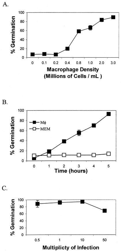
Effects of Mφ density, time, and MOI on the germination of B. anthracis Sterne. (A) Effect of Mφ cell density. RAW 264.7 Mφ (0 to 3 × 10⁶/ml) in MEM were added to wells of a 24-well tissue culture plate and infected with 106 endospores in a final volume of 1 ml. The plates were incubated at 37°C with 5% CO2 for 60 min. After 60 min, the percentage of endospore germination was calculated as described in the text. (B) Time course. A subgerminal concentration of RAW 264.7 Mφ (0.2 × 10⁶/ml) was infected at an MOI of 5:1 with Sterne endospores to a final endospore concentration of 106/ml (black squares). The Mφ were incubated at 37°C with 5% CO2, and measurements of the percentage of germination of radiolabeled endospores were taken hourly for 5 h. Germination assays performed with MEM alone served as a negative control (white squares). (C) Effect of MOI. The effect of the MOI on endospore germination was measured by using RAW 264.7 Mφ (2 × 10⁶/ml) and different concentrations of endospores. The Mφ were incubated at 37°C with 5% CO2 for 60 min. After 60 min, the percentage of endospore germination was calculated as described in the text. Each experiment was performed in triplicate with at least two separate preparations of labeled endospores, and results are reported with an error of ±1 standard deviation of the mean.
Macrophage cell culture.
RAW 264.7 murine (ATCC TIB-71) Mφ-like cells and J774A.1 (ATCC TIB-67) Mφ-like cells were cultured in Dulbecco's modified Eagle's medium with 10% fetal bovine serum (Gibco BRL) at 37°C with 5% CO2 in a humidified incubator. For germination assays, Mφ were washed twice with prewarmed serum-free minimal essential medium (MEM) or Hanks balanced salt solution (Hanks), resuspended in MEM or Hanks, and counted with a hemocytometer (Hausser Scientific). For cytochalasin D (Sigma) assays, Mφs were washed twice with MEM plus cytochalasin D (2 μg/ml) prior to being resuspended in fresh MEM plus cytochalasin D (2 μg/ml) with a final Mφ concentration of 2 × 10⁶ cells per ml. Conditioned medium was obtained at desired times by incubating Mφ in appropriate experimental media in six-well tissue culture plates (Costar) at 37°C with 5% CO2.
RESULTS
B. anthracis gerH is required for Mφ-related endospore germination.
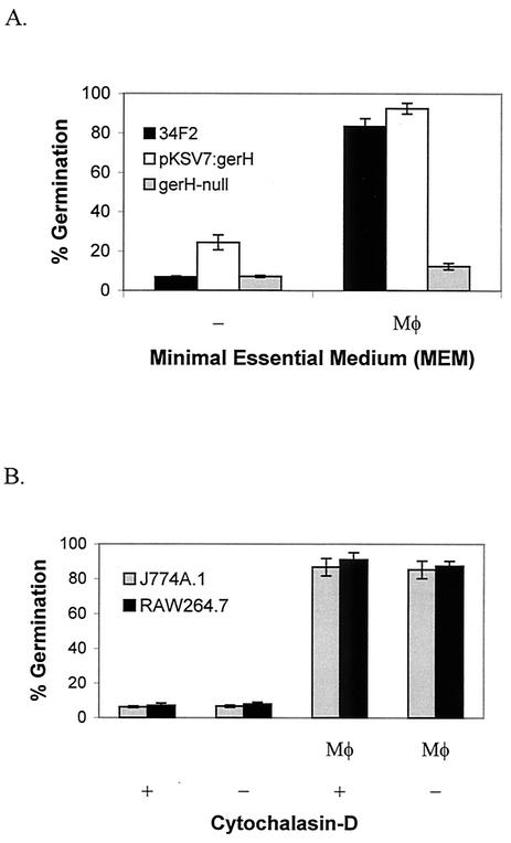
Differences between the germination of parental endospores and that of gerH-null endospores in Mφ cultures establish the degree of importance of gerH in cell-associated germination. B. anthracis Sterne parental endospores germinated completely in 60 min in RAW 264.7 Mφ cultures but not in MEM alone (Fig. 1). Conversely, gerHa-null endospores did not germinate in either Mφ culture or in MEM alone (Fig. 1A). Germination of the null strain in Mφ culture was rescued by complementation with wild-type gerHabc expressed under the control of its endogenous promoter from plasmid pKSV7 (Fig. 1A).
FIG. 1.
B. anthracis Sterne endospores require gerH for germination in macrophage cultures. (A) RAW 264.7 Mφ were infected with 45Ca-radiolabeled B. anthracis Sterne 34F2 (black bars), the gerH-null strain carrying pKSV7:gerH (open bars), and gerH-null endospores (grey bars). (B) Germination experiments were performed with 45Ca-radiolabeled 34F2 endospores in cultures of J774A.1 (grey bars) and RAW 264.7 (black bars) Mφ that were incubated in MEM with or without the addition of cytochalasin D. For all experiments, Mφ were infected with endospores and incubated in MEM at 37°C with 5% CO2 for 60 min. After 60 min, the percentage of endospore germination was calculated as described in the text. Each experiment was performed in triplicate with at least two separate preparations of labeled endospores, and results are reported with an error of ±1 standard deviation of the mean.
Treatment of Mφ cultures with cytochalasin D prior to in vitro infections with endospores inhibits phagocytosis and facilitates the study of extracellular germination in the presence of Mφ. Cytochalasin D inhibits actin polymerization and, therefore, any actin-dependent vesicle transport in eukaryotic cells (including phagocytosis). In studies with cytochalasin D, the extracellular germination of endospores suggested that Mφ secrete some germinants by actin-independent mechanisms. These studies were restricted to extracellular germination because no endospores were taken up by Mφ under these experimental conditions. Findings indicate that parental endospores germinated completely and equally in cultures of both J774A.1 and RAW 264.7 Mφ, with or without cytochalasin D (Fig. 1B). As predicted, no germination occurred in MEM alone, with or without cytochalasin D (Fig. 1B). Since MEM did not trigger germination of B. anthracis endospores, germination resulted from the conditioning of the MEM by Mφ. Because cytochalasin D broadly inhibits actin polymerization, we conclude that the conditioning of the extracellular medium by the Mφ occurred via actin-independent pathways.
Complete conditioning of the extracellular medium is dependent on both Mφ cell density and time.
The manner by which Mφ condition their extracellular environment with germinants was investigated by using different concentrations of Mφ and by performing a kinetic analysis of endospore germination in Mφ cultures. Germination experiments with 106 parental endospores/ml and RAW 264.7 Mφ densities ranging from 0.1 × 10⁶ to 3 × 10⁶/ml revealed a concentration-dependent germination response. A minimum of 0.4 × 10⁶ Mφ/ml was required to trigger germination above baseline in 60 min (Fig. 2A). A Mφ density of 3 × 10⁶/ml triggered the germination of 93% of the parental endospores in 60 min (Fig. 2A). After several hours, the majority of endospores could be observed floating freely in the extracellular medium, as endospores do not settle out of solution over the time course studied without centrifugation (data not shown). Thus, these experiments included extracellular germination, even without the addition of cytochalasin D to the Mφ cultures.
An investigation of the kinetics of germination in Mφ cultures revealed a time-dependent conditioning of the extracellular medium. If a low, initially subgerminal, density of Mφ acquires with time the ability to trigger germination of endospores, then it is possible that Mφ secrete active germinants into the medium. We found that an initially subgerminal Mφ cell density of 0.2 × 10⁶/ml (Fig. 2A) became capable of conditioning the medium with time (Fig. 2B). Additionally, a time course of germination with parental endospores determined with a Mφ density of 0.2 × 10⁶/ml indicated that germination above baseline begins after 2 h and reaches 93% after 5 h of incubation. In cultures with a Mφ density of 2 × 10⁶/ml, the total percentage of germination was unchanged when the multiplicity of infection (MOI) was increased from 0.5 (106 endospores/ml) to 10 and decreased only slightly at an MOI of 50 (Fig. 2C). This finding indicates that there are abundant levels of germinants in conditioned medium and that endospore density does not significantly affect germination at the concentrations tested. Together, the data in Fig. 2 demonstrated that Mφ condition media steadily over time, that higher densities of Mφ will condition media faster than lower densities, and that the ability of endospores to germinate in conditioned media does not relate to the MOI used in the experiment.
Evidence that Mφ secrete a purine required for gerH-mediated germination.
Our initial report (25) on gerH characterized the profile of the binary pairs of cogerminant molecules that trigger in vitro germination of the gerH-null strain relative to that of the parental Sterne strain of B. anthracis. That report described the Ino-His germination response of B. anthracis, which requires gerH, in which neither adenosine nor guanosine can substitute for Ino (25). In contrast, Ino can be replaced by adenosine and guanosine in the Ino-Ala germination response pathway. Therefore, Ino-His and Ino-Ala represent distinct germination pathways (25). Additionally, germination of B. anthracis endospores can occur in l-Ala alone at concentrations of 100 mM in water and in l-Ala at concentrations as low as 1 mM in certain salt solutions (11, 25) (Fig. 3A). Our finding that gerH is required for germination in Mφ cultures suggests an important role for the Ino-His germination response pathway. If Mφ secrete purines or amino acids that participate in the Ino-His germination response pathway, then the addition of a complementary exogenous cogerminant should trigger germination. Because amino acid and nucleoside cogerminant profiles of B. anthracis are known, we may then infer the nature of Mφ-secreted germinants from the identity of the defined cogerminant added to the solution.
FIG. 3.
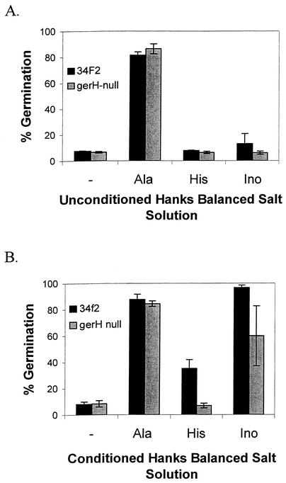
Partial characterization of Mφ-secreted germinants in Hanks with B. anthracis Sterne (black bars) and gerH-null endospores (grey bars). (A) Hanks was used alone or was supplemented with 1 mM Ala, 100 mM His, or 100 mM Ino. (B) Hanks was conditioned with 2 × 10⁶ Mφ/ml for 60 min and then passed through a sterile filter as described in the text. Ala, His, or Ino was added to conditioned Hanks at the concentrations listed above. Radiolabeled endospores were added to a final concentration of 106 endospores/ml and were incubated at room temperature for 60 min. After 60 min, the percentage of endospore germination was calculated as described in the text. Each experiment was performed in triplicate with at least two separate preparations of labeled endospores, and the results are reported with an error of ±1 standard deviation of the mean.
To characterize germinants secreted by RAW 264.7 Mφ, cells were washed thoroughly with Hanks and incubated in fresh Hanks for 3 h to allow Mφ conditioning of this buffer, which was then passed through a sterile filter and used in germination assays (as described above). Hanks is a salt solution that does not contain any germinant molecules. Unconditioned Hanks or conditioned Hanks alone did not trigger endospore germination, while the addition of 1 mM l-alanine to either solution did, as expected (Fig. 3). Germination studies of parental and gerH-null endospores performed with conditioned Hanks supplemented with histidine or Ino (100 and 1 mM, respectively) identified cogerminants that pair effectively with Mφ-secreted germinants. Conditioned Hanks, supplemented with His, triggered the germination of parental endospores in 60 min (Fig. 3B), in contrast to unconditioned Hanks with His, which did not (Fig. 3A). His participates in two distinct germination pathways: Ino-His and Ala-His (11, 25). Conditioned Hanks, supplemented with His, did not trigger germination in gerH-null endospores (Fig. 3B). gerH-null endospores maintain an intact Ala-His germination response but lack the Ino-His germination response (25). We can conclude from these data that Ino is likely to be one of the cogerminants secreted by Mφ because of the inability of other purines to substitute for Ino in the Ino-His germination pathway (25). Additionally, since purines act as cogerminants for B. anthracis endospores only in the presence of specific amino acids (11, 25), germination in Hanks supplemented with Ino would indicate the secretion of at least one amino acid by Mφ. However, the possibility also exists that the secreted molecules may be novel germinants that have yet to be identified. We report here that conditioned Hanks supplemented with Ino triggered germination in both parental and gerH-null endospores, while unconditioned Hanks supplemented with Ino did not (Fig. 3). These data strongly suggest that Mφ also secrete, along with Ino, at least some amino acids into the extracellular medium.
By using different cogerminants of B. anthracis endospores that trigger germination only when in binary combination with a specific cogerminant, we were able to report here that Mφ secrete a purine, which is likely Ino, and at least one or more of several candidate amino acids. The decrease of approximately 40% in the germination of gerH-null endospores relative to that of the parental endospores in conditioned Hanks with Ino is likely to be due to the absence of the Ino-His germination response pathway controlled by the gerH operon (Fig. 3B). The lack of germination in conditioned Hanks alone suggests that the concentrations of the secreted cogerminants are too low under the experimental conditions measured to trigger germination or that the correct binary pairs of cogerminants do not exist in the secreted medium. Because we used endospore germination in conditioned Hanks as our measure of a secreted cogerminant, these findings suggest a functional role for Ino and the other cogerminants in vivo that may be sensed by gerH-encoded proteins.
DISCUSSION
The absence of germination of B. anthracis gerH-null endospores in Mφ cultures and in Mφ-conditioned medium demonstrates that the germination operon gerH is required for Mφ-mediated germination. Mφ secrete one or more cogerminants that fulfill the role that Ino plays in vitro, with respect to gerH-dependent germination, in the presence of His. The gerH-dependent Ino-His germination response is absent in gerH-null endospores (25). This absence is likely responsible for the loss of the ability of the mutant to germinate in Mφ cultures and in Mφ-conditioned medium. Mφ may also contribute amino acids to the extracellular medium in sufficiently high concentrations to perform as cogerminants when exogenous Ino is added to the medium. The data reported here argue against the theory that Mφ condition the extracellular medium by consuming inhibitors of germination because it is highly unlikely that Hanks contains any molecular species that can affect endospore germination.
Several previous studies implicate alveolar Mφ as the de facto sites of B. anthracis endospore germination although, in vitro, other cell types can fulfill this role (3, 6, 20). While B. anthracis endospore germination appears to occur inside of Mφ in vivo, it remains unclear whether this process can occur extracellularly in an animal. The data reported here suggest that B. anthracis endospores may be able to germinate extracellularly in vivo in a position proximal to phagocytes prior to their uptake. Endospores can germinate extracellularly in vitro in MEM conditioned by Mφ. Electron micrographs created by Dixon et al. and the work of Welkos et al. have recently shown both intact endospores and recently germinated endospores within Mφ phagosomes (3, 26, 27, 28). Welkos et al. reported a Mφ sporicidal activity associated with RAW 264.7 cells that we did not observe. The gerH-null strain may be useful for addressing the specific question of sporicidal activity since these endospores do not germinate in the presence of Mφ. The 45Ca radiolabel is retained by these endospores, which indicates that they are structurally intact, and serves as a marker for viability. These endospores can be recovered and grown on BHI agar. It is possible that the sporicidal activity reported previously is the result of endospores germinating immediately prior to being taken up by the Mφ, which then kills a nascent vegetative cell and not an intact endospore.
It is becoming increasingly apparent that B. anthracis endospore germination is a multifactorial event. Like gerHabc, the germination operon gerSabc is also required for Mφ-related germination (12). The addition of d-alanine, a potent inhibitor of the Ala germination response, also inhibits germination in Mφ cultures (data not shown) and implicates Ala in Mφ-mediated germination. In other studies, the disruption of the pXO1-carried gerX partially attenuates endospore germination in Mφ (6), although ΔSterne endospores lacking pXO1 respond to all known germinants and are fully capable of germinating in Mφ cultures and in mice (11, 26, 27, 28). Collectively, these studies suggest that B. anthracis endospores rely on multiple chromosomally encoded ger operons to recognize complex patterns of discrete chemical cogerminants at low concentrations. While the Ino-His germination pathway is required for Mφ-related germination, this path is likely to be only one of a variety of germination response pathways contributing to Mφ-related germination. Therefore, the term “Mφ-mediated germination” may refer to germination in the presence of a variety of germinants secreted by host cells in low concentrations rather than to germination in response to germinants provided only by Mφ and not by any other source.
Acknowledgments
We thank B. Thomason, S. Cendrowski, B. Heffernan, N. Fisher, J. Crossno, and N. Bergman for their comments on this work.
This work was supported in part by NIH grants AI-08649 and AI-40644 and by ONR grants N00014-00-1-0422, N00014-01-1-1044, and N00014-02-1-0061.
Editor: D. L. Burns
REFERENCES
- 1.Barlass, P. J., C. W. Huston, M. O. Clements, and A. Moir. 2002. Germination of Bacillus cereus spores in response to l-alanine and to inosine: the roles of gerL and gerQ. Microbiology 148:2089-2095. [DOI] [PubMed]
- 2.Clements, M. O., and A. Moir. 1998. Role of the gerI operon of Bacillus cereus 569 in the response of spores to germinants. J. Bacteriol. 180:6729-6735. [DOI] [PMC free article] [PubMed] [Google Scholar]
- 3.Dixon, T. C., A. A. Fadl, T. M. Koehler, J. A. Swanson, and P. C. Hanna. 2000. Early Bacillus anthracis-macrophage interactions: intracellular survival and escape. Cell. Microbiol. 2:453-463. [DOI] [PubMed] [Google Scholar]
- 4.Dixon, T. C., M. Meselson, J. Guillemin, and P. C. Hanna. 1999. Anthrax. N. Engl. J. Med. 341:815-826. [DOI] [PubMed] [Google Scholar]
- 5.Foster, S. J., and K. Johnstone. 1990. Pulling the trigger: the mechanism of bacterial spore germination. Mol. Microbiol. 4:137-141. [DOI] [PubMed] [Google Scholar]
- 6.Guidi-Rontani, C., Y. Pereira, S. Ruffie, J. C. Sirard, M. Wever-Levy, and M. Mock. 1999. Identification and characterization of a germination operon on the virulence plasmid pXO1 of Bacillus anthracis. Mol. Microbiol. 33:407-414. [DOI] [PubMed] [Google Scholar]
- 7.Hachisuka, Y. 1969. Germination of B. anthracis spores in peritoneal cavity of rats and establishment of anthrax. Jpn. J. Microbiol. 13:199-207. [DOI] [PubMed] [Google Scholar]
- 8.Hanna, P. C., and J. A. Ireland. 1999. Understanding Bacillus anthracis pathogenesis. Trends Microbiol. 7:180-182. [DOI] [PubMed] [Google Scholar]
- 9.Hills, G. M. 1949. Chemical factors in the germination of spore bearing aerobes. The effects of amino acids on the germination of Bacillus anthracis, with some observations on the relation of optical form to biological activity. Biochem. J. 45:363-370. [DOI] [PMC free article] [PubMed] [Google Scholar]
- 10.Hudson, K. D., B. M. Corfe, E. H. Kemp, I. M. Feavers, P. J. Coote, and A. Moir. 2001. Localization of GerAA and GerAC germination proteins in the Bacillus subtilis spore. J. Bacteriol. 183:4317-4322. [DOI] [PMC free article] [PubMed] [Google Scholar]
- 11.Ireland, J. A. W., and P. C. Hanna. 2002. Amino acid-and purine ribonucleoside-induced germination of Bacillus anthracis ΔSterne endospores: gerS mediates responses to aromatic ring structures. J. Bacteriol. 184:1296-1303. [DOI] [PMC free article] [PubMed] [Google Scholar]
- 12.Ireland, J. A. W., and P. Hanna. 2002. Macrophage-enhanced germination of Bacillus anthracis endospores requires gerS. Infect. Immun. 70:5870-5872. [DOI] [PMC free article] [PubMed] [Google Scholar]
- 13.Kim, H. U., and J. M. Goepfort. 1974. A sporulation medium for Bacillus anthracis. J. Appl. Bacteriol. 37:265-267. [DOI] [PubMed] [Google Scholar]
- 14.Koehler, T. M., Z. Dai, and M. Kaufman-Yarbray. 1994. Regulation of the Bacillus anthracis protective antigen gene: CO2 and a trans-acting element activate transcription from one of two promoters. J. Bacteriol. 176:586-595. [DOI] [PMC free article] [PubMed] [Google Scholar]
- 15.McCann, K. P., C. Robinson, R. L. Sammons, D. A. Smith, and B. M. Corfe. 1996. Alanine germination receptors of Bacillus subtilis. Lett. Appl. Microbiol. 23:290-294. [DOI] [PubMed] [Google Scholar]
- 16.Moir, A., E. H. Kemp, C. Robinson, and B. M. Corfe. 1994. The genetic analysis of bacterial spore germination. J. Appl. Bacteriol. 77:9S-16S. [PubMed] [Google Scholar]
- 17.Moir, A., B. M. Corfe, and J Behraven. 2002. Spore germination. Cell. Mol. Life Sci. 59:403-409. [DOI] [PMC free article] [PubMed] [Google Scholar]
- 18.Paidhungat, M., and P. Setlow. 1999. Isolation and characterization of mutations in Bacillus subtilis that allow spore germination in the novel germinant d-alanine. J. Bacteriol. 181:3341-3350. [DOI] [PMC free article] [PubMed] [Google Scholar]
- 19.Paidhungat, M., and P. Setlow. 2000. Role of Ger proteins in nutrient and nonnutrient triggering of spore germination in Bacillus subtilis. J. Bacteriol. 182:2513-2519. [DOI] [PMC free article] [PubMed] [Google Scholar]
- 20.Ross, J. M. 1957. Pathogenesis of anthrax following administration of spores by the respiratory route. J. Pathol. Bacteriol. 73:485-494. [Google Scholar]
- 21.Smith, K., and P. Youngman. 1992. Use of a new integrated vector to investigate compartment specific expression of the Bacillus subtilis spoIIM gene. Biochimie 74:705-711. [DOI] [PubMed] [Google Scholar]
- 22.Stewart, G., K. Johnstone, E. Hagelberg, and D. J. Ellar. 1981. Commitment of bacterial spores to germinate. A measure of the trigger reaction. Biochem. J. 198:101-106. [DOI] [PMC free article] [PubMed] [Google Scholar]
- 23.Suzuki, J. B., R. Booth, and N. Grecz. 1971. In vivo and in vitro release of Ca [45] from spores of Clostridium botulinum type A as further evidence for spore germination. Res. Commun. Chem. Pathol. Pharmacol. 2:16-23. [PubMed] [Google Scholar]
- 24.Titball, R. W., and R. J. Manchee. 1987. Factors affecting the germination of spores of Bacillus anthracis. J. Appl. Bacteriol. 62:269-273. [DOI] [PubMed] [Google Scholar]
- 25.Weiner, M. A., T. D. Read, and P. C. Hanna. 2003. Identification and characterization of the gerH operon of Bacillus anthracis endospores: a differential role for purine nucleosides in germination. J. Bacteriol. 185:1462-1464. [DOI] [PMC free article] [PubMed] [Google Scholar]
- 26.Welkos, S. L., R. W. Trotter, D. M. Becker, and G. O. Nelson. 1989. Resistance to the Sterne strain of B. anthracis: phagocytic cell responses of resistant and susceptible mice. Microb. Pathog. 7:15-35. [DOI] [PubMed] [Google Scholar]
- 27.Welkos, S., S. Little, A. Friedlander, D. Fritz, and P. Fellows. 2001. The role of antibodies to Bacillus anthracis and anthrax toxin components in inhibiting the early stages of infection by anthrax spores. Microbiology 147:1677-1685. [DOI] [PubMed] [Google Scholar]
- 28.Welkos, S., A. Friedlander, S. Weeks, S. Little, and I. Mendelson. 2002. In-vitro characterisation of the phagocytosis and fate of anthrax spores in macrophages and the effects and anti-PA antibody. J. Med. Microbiol. 51:821-831. [DOI] [PubMed] [Google Scholar]
- 29.Yosuda, Y., S. Yoshihiro, and K. Tockikubo. 1996. Immunological detection of the GerA spore germination proteins in the spore integuments of Bacillus subtilis using scanning electron microscopy. FEMS Microbiol. Lett. 139:235-238. [DOI] [PubMed] [Google Scholar]
- 30.Zuberi, A. R., I. M. Feavers, and A. Moir. 1985. Identification of three complementation units in the gerA spore germination locus of Bacillus subtilis. J. Bacteriol. 162:756-762. [DOI] [PMC free article] [PubMed] [Google Scholar]


