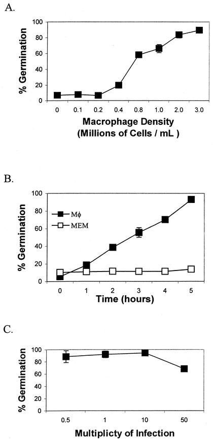
FIG. 2.
Effects of Mφ density, time, and MOI on the germination of B. anthracis Sterne. (A) Effect of Mφ cell density. RAW 264.7 Mφ (0 to 3 × 10⁶/ml) in MEM were added to wells of a 24-well tissue culture plate and infected with 106 endospores in a final volume of 1 ml. The plates were incubated at 37°C with 5% CO2 for 60 min. After 60 min, the percentage of endospore germination was calculated as described in the text. (B) Time course. A subgerminal concentration of RAW 264.7 Mφ (0.2 × 10⁶/ml) was infected at an MOI of 5:1 with Sterne endospores to a final endospore concentration of 106/ml (black squares). The Mφ were incubated at 37°C with 5% CO2, and measurements of the percentage of germination of radiolabeled endospores were taken hourly for 5 h. Germination assays performed with MEM alone served as a negative control (white squares). (C) Effect of MOI. The effect of the MOI on endospore germination was measured by using RAW 264.7 Mφ (2 × 10⁶/ml) and different concentrations of endospores. The Mφ were incubated at 37°C with 5% CO2 for 60 min. After 60 min, the percentage of endospore germination was calculated as described in the text. Each experiment was performed in triplicate with at least two separate preparations of labeled endospores, and results are reported with an error of ±1 standard deviation of the mean.

