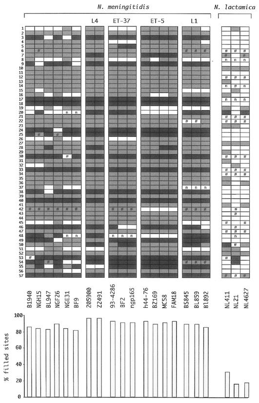
FIG. 3.
Conservation of nemis repeats in neisserial chromosomes. The distribution of the 57 nemis elements listed in Fig. 2 is diagrammed as follows: empty and filled boxes represent intergenic chromosomal regions lacking and containing nemis elements, respectively. The presence of long and short nemis is marked by light and dark grey filling, respectively. The number sign represents regions differing in size from either filled or empty sites. Regions for which reliable PCR amplification signals could not be obtained, regardless of changes in either PCR settings or primer pairs, are labeled by n. The relative abundance of nemis-positive regions within each strain is highlighted in the histogram at the bottom.

