
Fig. 1.
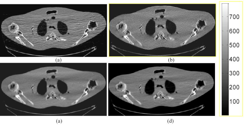
Shoulder phantom study with 10 mA protocol. (a): A conventional FBP reconstructed image using the Hanning filter with cutoff at 80% Nyquist frequency. (b): Iterative PRWLS+SOR reconstructed image with β =1×10⁻⁴. (c): A standard FBP reconstructed image from the iterative GS-PRWLS smoothed sinogram with β =1×10⁻⁸. (d): A standard FBP reconstructed image from the analytical KL-PWLS filtered sinogram with β =200.