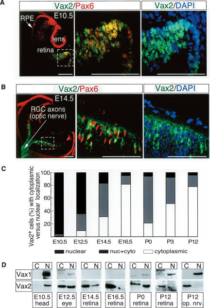
Figure 1.
Dynamic nuclear–cytoplasmic translocation of Vax2 during retinal development. (A,B) Ten-micrometer sections of E10.5 (A) and E14.5 (B) embryonic mouse eyes costained with a rabbit polyclonal Vax2 antibody (green) and a mouse monoclonal Pax6 antibody (red). Vax2 and Pax6 are coexpressed in the E10.5 ventral optic cup (A) and E14.5 ventral retina (B). Both proteins are localized to nuclei at E10.5, whereas Vax2 is prominent in the cytoplasm and axons of RGCs at E14.5 (shown in B). Left and middle panels of A and B are Vax2/Pax6 coimmunostaining, with the middle panels corresponding to enlarged images of the boxed area in the left panels. Right panels are equivalent to the middle panels, and illustrate Vax2 immunostaining together with DAPI staining (blue) to visualize nuclei. Bars, 100 μm. (C) Quantitation of the fraction of Vax2+ retinal cells displaying nuclear versus cytoplasmic localization of the protein, as a function of time in development. Confocal images of Vax2 immunostaining from retinal sections were counted for cells expressing Vax2 exclusively in the cytoplasm (open bars), in both the nucleus and the cytoplasm (gray bars), or exclusively in the nucleus (black bars). Numbers of cells in each category were divided by the total Vax2+ cells counted to obtain the percentage values displayed in the graph. Values are the mean from five sections (>200 total cells counted per section). (D) Homogenates of the indicated tissues were separated into nuclear and cytoplasmic fractions (see Materials and Methods) and monitored for Vax1 or Vax2 protein expression by Western blot with anti-Vax1 or anti-Vax2 antibodies.

