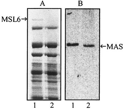
FIG. 2.
SDS-PAGE (A) and immunoblot (B) analyses of total extracts from wild-type and msl6 mutant M. tuberculosis. The separated proteins were stained with Coomassie blue or analyzed by immunoblotting with anti-MAS antibodies. Lane 1, wild type; lane 2, msl6 mutant. The protein band at 430 kDa (MSL6) was used for amino acid sequence analysis.

