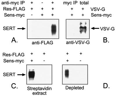
Figure 4.
Co-precipitation of Res-FLAG with Sens-myc. (A) HeLa cells transfected with equal amounts of Res-FLAG and Sens-myc cDNA, or with Res-FLAG alone, were solubilized and treated with Protein-A beads and, where indicated, antibody against c-myc. The immunoprecipitates were separated by SDS/PAGE and were blotted with anti-FLAG antibody as described. (B) Cells expressing Sens-myc and VSV-G protein were treated as in A. The immunoprecipitate in the left lane is compared with the initial cell lysate in the right lane. (C) Sens-myc on the surface of cells expressing Res-FLAG and Sens-myc, or Res-FLAG alone, was labeled with 1 mM MTSEA-biotin, solubilized, precipitated with streptavidin-agarose, and Western blotted with anti-FLAG antibody. (D) The remaining soluble extract after depletion of cell lysate with MTSEA-biotin, and streptavidin-agarose, as in C, was immunoprecipitated and Western blotted as in A. Although the sample in C represents quantitative precipitation of biotinylated proteins, the fraction of intracellular SERT precipitated with anti-myc antibodies (D) was not determined.

