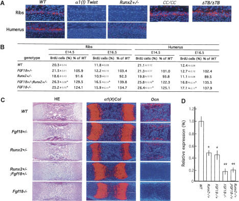
Figure 3.
Runx2 regulates chondrocyte proliferation through Fgf18. (A) In situ hybridization analysis of Fgf18 expression in ribs and humeri of E15.5 wild-type (WT), α1(I) Twist-1, Runx2 +/−, CC/CC, and ΔTwist box −/− embryos. Perichondrial Fgf18 expression is decreased in α1(I) Twist1 and Runx2 +/− and increased in CC/CC and ΔTwist box −/− embryos. (B) BrdU incorporation analysis in ribs and humeri at E14.5 and E16.5 of wild-type, Fgf18 +/−, Runx2 +/−, Fgf18 +/− ; Runx2 +/−, and Fgf18 −/− embryos. Chondrocyte proliferation is similarly increased in Fgf18 +/− ; Runx2 +/− and Fgf18 −/− embryos. (C) Histological and in situ hybridization analysis of α1(X) Collagen and Osteocalcin expression in humeri of E16.5 wild-type, Fgf18 +/−, Runx2 +/−, Fgf18 +/− ; Runx2 +/−, and Fgf18 −/− embryos. (D) Real-time PCR analysis of α1 Integrin expression in humeri of E15.5 wild-type, Runx2 +/−, Fgf18 +/−, Fgf18 −/−, and Runx2 +/− ; Fgf18 +/− embryos, normalized to β-actin.

