Figure 2.
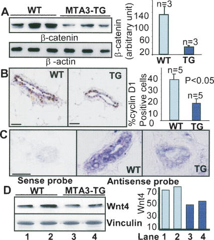
Impaired Wnt pathway in the MTA3-TG mammary tissue. (A) Western blot analysis of β-catenin in the virgin mammary tissue. (B) IHC analysis of cyclin D1 in the wild-type (WT) and TG mammary tissues. Bars, 100 μm. (C) In situ hybridization of digoxigenin-labeled Wnt4 cDNA antisense transcripts to mammary tissue sections. Bars, 100 μm. (D) Western blot analysis of Wnt4 from tissue lysates. Mammary glands used were from the virgin 12-wk-old female mice.

