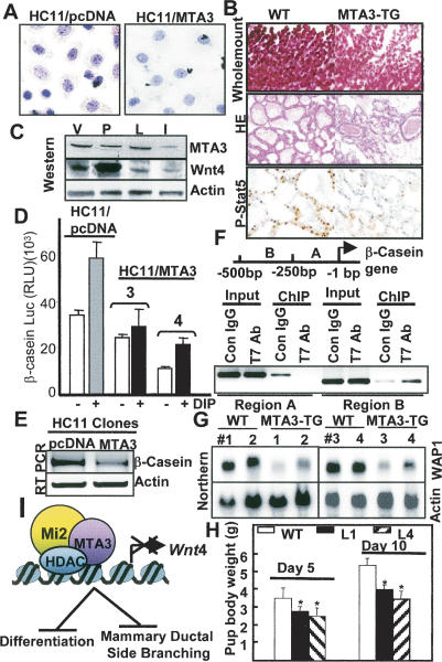
Figure 5.
MTA3 suppresses mammary gland differentiation and morphogenesis. (A) Status of the oil red-O-stained neutral lipid droplets in the HC11 clones treated with DIP for 3 d. (B) Whole-mount staining, H&E staining, and immunostaining for P-STAT5 of mammary tissues at lactation day 2. (C) Status of Wnt4 and MTA3 proteins in various stages of the wild-type mammary gland development. (V) Virgin day (12 wk); (P) pregnancy day 4; (L) lactation day 1; (I) involution day 5. (D) Repression of β-casein-luc activity in HC11/ MTA3 cells (E) Repression of the levels of β-casein mRNA in HC11/ MTA3 cells in comparison with the levels in HC11/pcDNA cells. (F) ChIP showing association of MTA3 with a 250-bp region of the β-casein promoter encompassing −250 bp to −500 bp. (G) Northern blot analysis of the WAP1 in mammary glands from the lactation day 2 wild-type (WT) and MTA3-TG mice. (H) Pup body weight at lactation days 5 and 10. Values are average weight ± standard deviation. P < 0.05 for L1 and L4 according to Student's t-test. (I) Schematic diagram of regulation of Wnt4 expression by the MTA3/NuRD complex and its consequences on mammary gland development.

