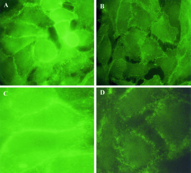
Figure 7.
Effect of PAF on the junctional localization of β-catenin and VE-cadherin in KS cells. Cells were grown to subconfluence and treated with 10 ng/ml of PAF for 2 hours. A and C represent control staining for β-catenin and VE-cadherin, respectively; B and D represent β-catenin and VE-cadherin staining after PAF treatment. In B and D, it is evident the gap formation and absence of β-catenin and VE-cadherin in areas where the cells have separated. PAF also promoted a zigzag pattern distribution of β-catenin (B) and a diffuse staining pattern for VE-cadherin (D) different from controls displaying a linear pattern distribution localized at cell-cell contact. Three experiments were performed with similar results. Original magnifications, ×400.

