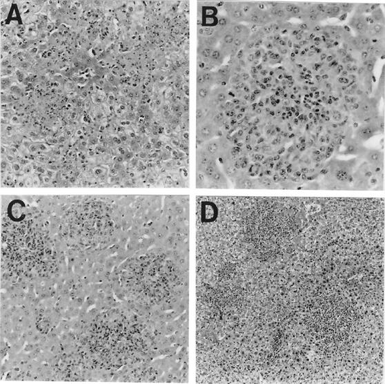
FIG. 2.
Characteristic histopathological changes in liver caused by Y. pestis KIM10 on postinfection day 3 (hematoxylin and eosin stain). (A) Control mouse infected with pCD+ yersiniae showing multiple necrotic focal lesions without an inflammatory cell response (magnification, ×140); (B) control mouse infected with pCD− yersiniae exhibiting granuloma formation (magnification, ×280); (C) mouse actively immunized with PAV and then infected with pCD+ yersiniae showing protective granulomatous lesions (magnification, ×140); (D) mouse passively immunized with polyclonal rabbit anti-PAV and infected with pCD+ yersiniae showing pregranulomatous lesions prompting infiltration of inflammatory (mononuclear) cells (magnification, ×70). This figure was reprinted from the work of Nakijima et al. (68).

